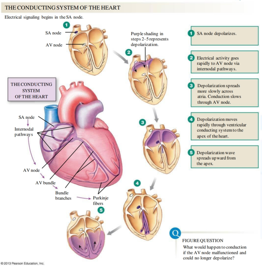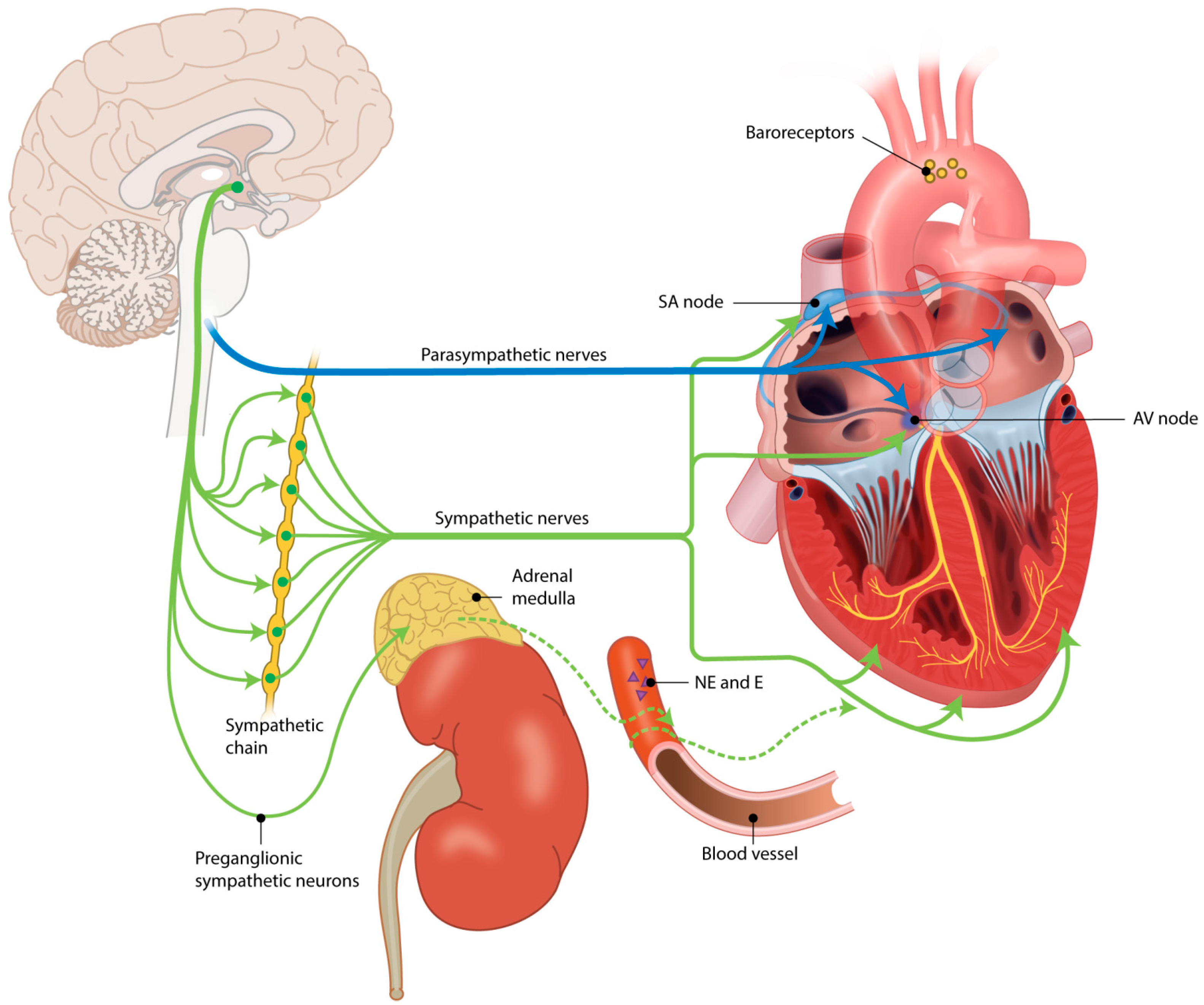

The current paradigm of SA node automaticity has been modeled as two clocks that function in concert, the “membrane voltage clock” and the “calcium clock”.

Pacemaker automaticity is due to spontaneous diastolic depolarization of phase 4, which depolarizes the membrane to threshold potential generating rhythmic action potentials. Peripheral SAN cells are electrophysiologically intermediate between central cells and atrial cardiomyocytes. The central SAN, the site of dominant pacemaking, is electrotonically insulated from the hyperpolarizing atrial myocardium through the differential expression of connexins and ion channels. Toxic mutant RNA and altered splicing factor functionĮlectrophysiologic heterogeneity of the sinoatrial node Myotonic dystrophy type I (Steinert’s disease)ĪV block, intraventricular conduction disease Sinus bradycardia and heart rate variabilityĪltered ion channel and transporter expression and traffickingĪltered ventricular myocyte energetics leading to glycogen engorged vacuoles and disruption of annulus fibrosus Specification defect of the AVN and the ventricular CCSĪltered nuclear stress mechanics and hyperactivation of MAPK signaling Sinus bradycardia, AV block, bundle branch block Impaired myocyte coupling resulting in slowed conduction Prolongation of action potential duration and reduced cardiac excitabilityĪltered Ca2+ handling likely affecting the Ca2+ clock Loss of depolarizing current in peripheral SAN cells

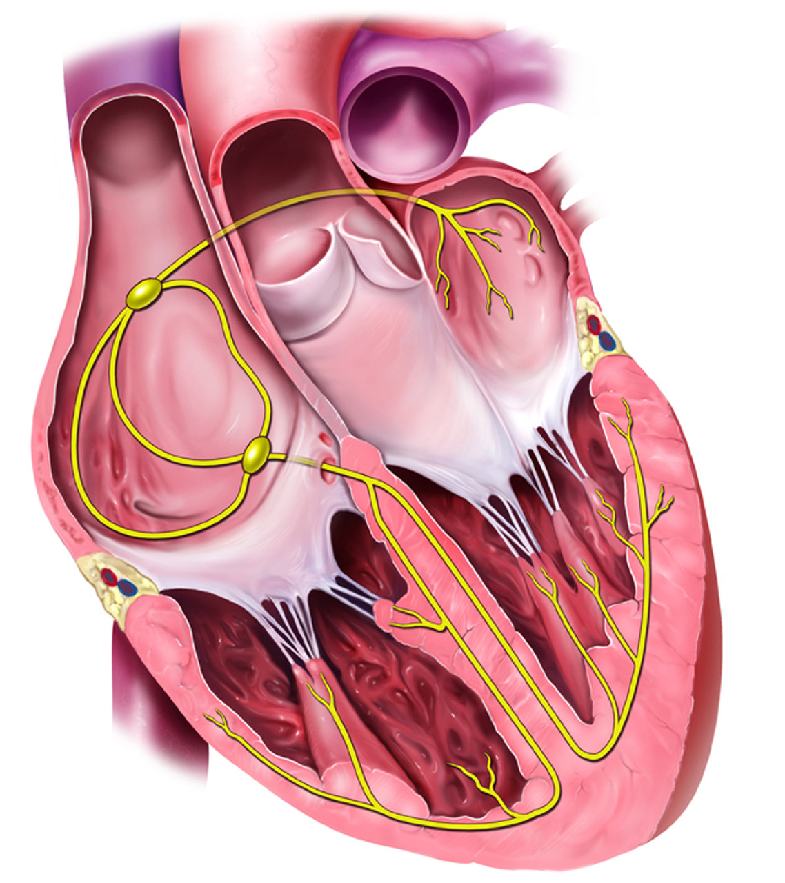
Reduced cardiac excitability and slowed conduction. SSS, PCCD, atrial standstill, AV block, bundle branch block We also investigate evolving therapeutic strategies that may serve as adjuvant or replacement therapy to current implantable pacemakers. In this review, we discuss gene families that have been implicated in human CCS diseases of rhythm, conduction block, accessory conduction, and development ( Table I). Applying a multidisciplinary approach, which includes human genetic screening, biophysical analysis, and transgenic mouse technology, has yielded a broad array of gene families involved in maintaining normal CCS physiology ( figure 1). Inherited forms of CCS disease are rare, but each new mutation provides invaluable insight into the molecular mechanisms governing CCS development and function. CCS dysfunction is primarily due to acquired conditions such as myocardial ischemia/infarct, age-related degeneration, procedural complications, and drug toxicity. Human diseases of the conduction system have been identified that alter impulse generation, propagation, or both. The functional components of the CCS can be broadly divided into the impulse generating nodes and the impulse propagating His-Purkinje system. The human heart beats 2.5 billion times during a normal lifespan, a feat accomplished by cells of the cardiac conduction system (CCS).


 0 kommentar(er)
